
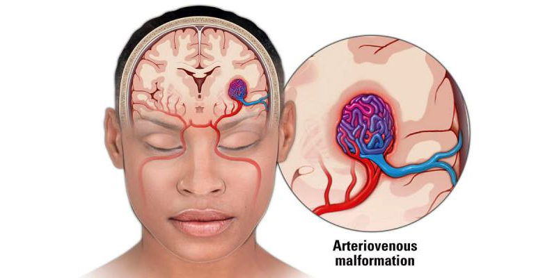
Arteriovenous malformation (AVM) surgeries are neurosurgical procedures aimed at treating abnormal connections between arteries and veins in the brain or spinal cord. AVMs pose a risk of bleeding, and surgical intervention may be considered to eliminate or reduce this risk. The specific approach to AVM surgery can vary based on the AVM's location, size, and characteristics. Here's an overview of common surgical options:
Microsurgical Resection:
Indication: Microsurgical resection involves removing the AVM using microsurgical techniques.
Procedure: A craniotomy is performed to expose the brain, and the surgeon uses a microscope for precision. The abnormal blood vessels are carefully dissected and removed. This approach is often considered for smaller, accessible AVMs.
Embolization:
Indication: Embolization is sometimes used as a preoperative or standalone treatment to reduce AVM size and blood flow.
Procedure: A catheter is threaded through the blood vessels to the AVM, and embolic agents (particles or glue) are injected to block or reduce blood flow. This may make the AVM more amenable to surgical removal or reduce the risk of bleeding.
Radiosurgery (Stereotactic Radiosurgery):
Indication: Radiosurgery is a non-invasive option suitable for certain AVMs, particularly those in locations difficult to access surgically.
Procedure: Precise beams of radiation are delivered to the AVM, causing changes over time that lead to the closure of abnormal vessels. This method is often chosen for smaller AVMs or those with higher surgical risks.
Combined Approaches:
Indication: Complex AVMs may require a combination of embolization, radiosurgery, and microsurgical resection.
Procedure: Treatment may be staged, with embolization performed first to reduce AVM size, followed by radiosurgery or surgical resection. The choice of approach is tailored to the individual case.
Endovascular Procedures:
Indication: In some cases, endovascular techniques may be used to treat certain aspects of an AVM.
Procedure: Using catheters and imaging guidance, interventions such as embolization or vessel occlusion may be performed selectively.
Postoperative Monitoring:
Indication: After surgical resection, patients are monitored for any signs of complications, such as bleeding or neurological deficits.
Procedure: Regular imaging studies and clinical assessments are conducted during the recovery period.
It's important to note that the decision to pursue surgery or another treatment modality depends on the individual characteristics of the AVM and the patient's overall health. The choice of intervention is typically made collaboratively by a multidisciplinary team, including neurosurgeons, interventional neuroradiologists, and radiation oncologists. The goal of AVM surgery is to eliminate the risk of bleeding and improve the patient's quality of life while minimizing potential complications.